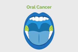
Oral cancer refers to cancers of the head and neck. It includes cancer of the lips, tongue, cheeks, gums, salivary glands, floor of the mouth, hard and soft palate, sinuses and pharynx. Brain cancer falls in a different category.

Dentist can identify any sign or abnormality during check-up and based on that they can refer Oral -maxillofacial surgeon - head and neck surgeon for further diagnosis.
- Examination of oral cavity, including lips, gums, tongue, cheeks, floor of the mouth, hard palate, soft palate, tonsillar area, buccal mucosa is done to check any abnormal changes such as red or white patches, lump, ulcers or lesions.
- Sub mandibular, sub lingual and cervical lymph nodes are examined to identify any swelling or pain.
Image 2 & 3 : Sub mandibular and cervical lymph node examination
- Colposcopy – It is a magnifying lens that helps doctor to have a close look at the mouth such as lesions, ulcers and areas of dysplasia. Biopsy is performed if any abnormality is identified.
- Optical diagnostic system – It is a non-invasive method used to detect early cancer. Changes in tissues and morphology are examined for early detection of cancer.
- VELscope - Fluorescence visualization, special light is used to detect changes in the oral mucosa.
- Toluidine blue (TB) and Lugols iodine staining – It is used to highlight the abnormal areas of mucosa. Abnormal tissue stains brown and normal tissue does not stain.
- Biopsy – There are various types of biopsies as mentioned below,
1. FNAC (fine needle aspiration cytology) – A long needle is inserted into the lesion to draw out the fluid and cells for examination.
2. Core needle biopsy – A larger needle with a cutting tip is inserted to take some tissues out from the suspected area.
3. Brush biopsy – A brush is used to collect the sample.
4. Incisional biopsy – A portion of the lump is removed.
5. Excisional biopsy – The whole organ or lump is removed.
6. Vacuum – assisted biopsy – A suction device is used to draw fluid and cells to take sample. - X-rays of the mouth: Radiographs (X-rays) check whether the cancer has extended into the bone.
- CT scan – It is used to detect primary tumours in the oral cavity. It is useful for staging oral cancer, determining the extent of tumour spread to nearby lymph nodes and distant organs.
- MRI – It is used to detect vascular invasion by the tumour, helping to determine the risk of metastasis and to take treatment decisions.
- PET – CT – In this test , a small amount of radioactive sugar is injected into patient. That is picked up by active cells like cancer cells. It is used to find oral cancer, spreading, invasion and analysing the treatment response.
For more information:
Changed
17/Jun/2025
Community
Condition


