
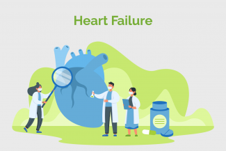
Heart Failure is a commonly seen condition amongst our aging population. In spite of advanced therapeutic science, incidence rates continue to increase. Life expectancy of patients with Heart Failure can be improved with lifestyle changes and compliance with their treatment plan. Mumbai-based cardiologist, Dr Nihar Mehta explains Heart Failure comprehensively.

How is Heart Failure diagnosed?
Heart failure is diagnosed based upon medical history, a physical examination, and a series of tests. The doctor will take into account the symptoms and risk factors and correlate those with clinical examination findings of heart rate, blood pressure, pressure wave forms of the venous pulse (JVP), chest and cardiac examination and examination of your abdomen and limbs These tests can determine how well your heart is working and cause of heart failure:
Electrocardiogram (ECG) - It is a commonly used diagnostic tool for heart failure.
- It measures the electrical activity of the heart through electrodes placed on the skin, recording the heart's rhythm and detecting any abnormalities. ECG findings can help differentiate heart failure from other conditions and guide further diagnostic testing. ECG may diagnose conditions that can cause heart failure such as abnormal heart rhythm or heart attack.
- In heart failure, an ECG may show specific changes indicating problems, such as enlarged heart chambers, thickening of the heart muscle, or irregular heart rhythms.
- Specific ECG abnormalities that may be seen in heart failure include prolonged QRS duration, electrical conduction delays, and changes in the ST segment or T wave.
Echocardiography – It is a non-invasive imaging technique that uses ultrasound to assess the structure and function of the heart in individuals with heart failure. It can help in identifying underlying cause of heart failure, such as coronary artery disease, valve disease or congenital heart defects.
- It provides real-time images of the heart, allowing for the evaluation of chamber sizes, wall thickness, and overall heart function.
- It measures the ejection fraction, which is the percentage of blood pumped out of the left ventricle with each heartbeat. This helps assess the heart's pumping ability. An ejection fraction of 50% or higher is considered ideal.
Blood tests – Complete blood count, thyroid function test, kidney function test, liver function test and lipid profile tests are done to detect underlying cause of heart failure.
- Basic Metabolic Panel (BMP) – It is a blood test that measures eight different chemicals that occur naturally in the blood to check electrolytes, fluid balance, glucose levels, and other markers of a person’s health. Carbon dioxide, sodium, potassium, and chloride. These electrically charged minerals help control the balance of acids and bases and the level of fluids in the body.
- Blood tests can measure specific biomarkers associated with heart failure.
- B type natriuretic peptide (BNP) – It is a hormone produced by the heart in response to increased pressure and stress, typically seen in heart failure. It is used to detect and access severity of heart failure. Normal BNP levels are less than 100 picograms per ml (pg/ml)
- N-terminal-proBNP (NT-proBNP) – It is released in response to cardiac stress and strain. It is done with BNP test to detect heart failure. For NT-proBNP, normal levels are less than 125 pg/mL.
- Galectin-3 – It is a protein associated with inflammation and fibrosis in cardiac diseases. Galectin-3 levels can provide insights into the prognosis and risk stratification of patients with heart failure. Optimal level for Galectin-3 is 0 to 22.2 ng/mL
- ST2 – It is another protein that is released in response to cardiac stress. Increased levels of soluble ST2 have been associated with adverse outcomes in heart failure.
- GDF-15: Growth Differentiation Factor-15 (GDF-15) is a protein involved in inflammation and cellular stress response. Elevated levels of GDF-15 have been found in heart failure patients and are associated with increased mortality risk.
- High sensitivity troponin: This is a more sensitive version of the traditional troponin biomarker, which is released when there is damage to the heart muscle. High sensitivity troponin tests can detect even small elevations, allowing for earlier detection of myocardial injury and potentially improving risk stratification in heart failure patients.
- Amyloid test: Abnormal deposition of Amyloid proteins in the heart can disrupt normal organ functioning. Tissue biopsy is done. Sample is taken from heart muscles. This test helps differentiate cardiac amyloidosis from other types of heart failure as it can have many underlying causes.
Chest X-ray – It shows the size and shape of heart and lung to detect any abnormality in size or shape. Chest X-ray helps detect the congestion or any accumulation of fluid in the lungs, which can sometimes be identified in individuals with heart failure.
Cardiac MRI – It helps assess cardiac function, measure ventricular volumes and provides detailed images of the heart structures and dysfunctional areas.
Cardiac CT scan - Computed tomography (CT) scans enable evaluation of the heart's arteries for blockages or narrowing. These scans help diagnose underlying causes of heart failure, such as coronary artery disease.
Exercise stress test - Patients perform physical exercise while monitoring heart function and symptoms. Advances in stress testing, such as cardiopulmonary exercise testing, can provide a comprehensive assessment of heart and lung function during exercise.
Changed
16/Mar/2025
Community
Condition