

Interstitial lung disease (ILD) is an umbrella term used for a large group of diseases that cause scarring (fibrosis) of the lungs. It affects millions of people globally. It is a serious condition that impacts mortality and quality of life of people affected by it.
ILD is different from COPD (Chronic Obstructive Pulmonary Disease)

What is Interstitial Lung Disease
To understand what Interstitial lung diseases actually are let’s try and understand what our lungs look like. Lungs are cylindrical organs, with a rounded, narrow peak at the top called the apex. At the base of the lungs is a broad, curved base that rests on the flattened surface of the diaphragm. The trachea (windpipe) divides into two main tubes called bronchus that divide several times after entering the lungs to form smaller tubes called bronchioles that eventually form the tiny air sacs called alveoli. The alveoli are where the gaseous exchange between the respiratory system and blood capillaries happens. From the hilum (an indent in the center of the lung), the lymphatic vessels, nerves, bronchus, and pulmonary arteries and veins enter the lungs.
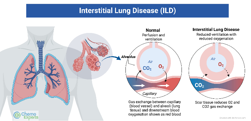
Image sourced from https://www.chemoexperts.com/interstitial-lung-disease.html
The interstitium pertains to the tissue region within and surrounding the wall of the air sacs (alveoli) in the lung area. It is where oxygen transfers from the alveoli to the capillary network (small blood vessels) that envelops the lung like a delicate layer of blood. Once the oxygen traverses the interstitial space, it enters the bloodstream and is transported to the vital organs of the body.
Interstitial lung diseases (ILDs) induce inflammation or scarring in this interstitial space, hindering the passage of oxygen into the bloodstream. This inflammation and scarring also stiffen the lung, increasing the effort required for breathing and causing a heightened sense of breathlessness, particularly during activities like climbing stairs along with other symptoms like a dry, persistent cough.
Changed
21/Apr/2025
Community
Condition


